Figure 3.
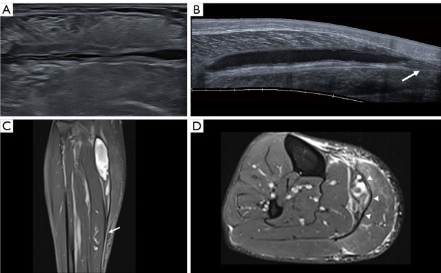
A 43-year-old man present a gastrocnemius aponeurotic injury (type 2B) at 5th day by US with a follow-up MRI at 3 weeks. He reported an acute injury while playing paddle and had to stop playing. Axial (A) and long (B) axis US images show the perimysium and muscle fascicles are retracted secondary to an aponeurotic rupture affecting more than 50% of total muscle width and a severe intermuscular hematoma. Free aponeurosis thick but without disruption (arrow). Sagittal/axial fat-suppressed TSE intermediate-weighted images (C,D) show the presence of reparative scar tissue along the anterior aponeurosis of the medial gastrocnemius muscle (arrowheads) with integrity of the free aponeurosis (arrow). See Video 3 showing asynchronous motion of the gastrocnemius and soleus muscles. US, ultrasound; MRI, magnetic resonance imaging; TSE, turbo spin-echo.

