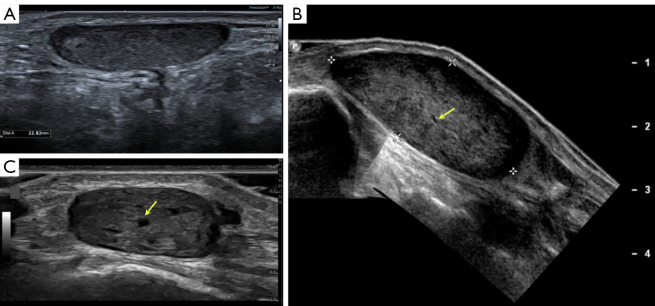
Figure 2.
B mode US typical angioleiomyoma pattern in three different patients: (A) female, 30 years old, longitudinal image of the left ankle; (B) male, 42 years old, longitudinal image of the ankle; (C) female, 52 years old, angioleiomyoma of the hand. All of them are subcutaneous solid and oval tumors, with smooth or lobulated well defined margins and a homogeneous and hypoechogenic background, showing small lineal anechoic images that correspond to small vessels (yellow arrows). US, ultrasonography.

