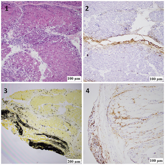
Figure 2.
(1) H&E staining of the frozen skeletal muscle block showing extensive areas of perifascicular atrophy, fiber damage, and perimysial mononuclear inflammation. (2) Alkaline phosphatase (ALP) staining of the frozen skeletal muscle block showing peripheral connective tissue reactivity adjacent to the areas of perifascicular abnormality and a significant increase in capillary uptake. (3) An anti-membrane attack complex c5b9 antibody frozen skeletal muscle block showing abnormal immunoreactivity of the perifascicular connective tissue adjacent to the areas of perifascicular abnormality and capillary uptake. (4) Anti-CD4 antibody, FFPE tissue block showing abundant T-helper lymphocytes in perifascicular areas of abnormality.

