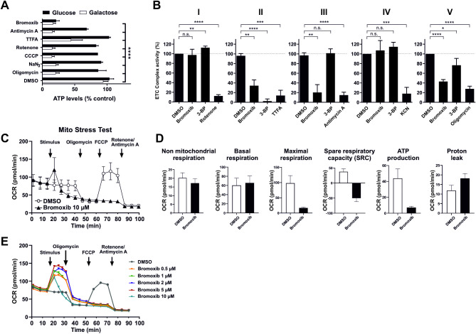
Fig. 5.
Bromoxib specifically targets mitochondrial metabolism, leading to the inhibition of both OXPHOS and glycolysis. (A) The impact of bromoxib (10 µM) and several known mitotoxins on the ATP levels in Ramos cells. The cells were treated with the specified agents in a complete growth medium containing either glucose or galactose as the sole available sugar. The following complex-specific inhibitors of the ETC were used: complex I: 10 µM rotenone; complex II: 10 µM thenoyltrifluoroacetone (TTFA); complex III: 10 µM antimycin A; complex IV: 1 mM sodium azide (NaN3); complex V: 10 µM oligomycin; mitochondrial uncoupler and protonophore: 1 µM CCCP and 10 µM bromoxib as well as DMSO (0.1% v/v) as vehicle control. ATP-levels were measured using the luminescence-based mitochondrial ToxGloTM assay (Promega). The depicted values were normalized to cells treated with DMSO in glucose containing growth medium (set to 100%). Error bars = mean ± SD of three independent experiments performed in triplicates; p-values were calculated by two-way ANOVA with the Holm-Sidak post-test; (**** = p ≤ 0.0001). (B) Bromoxib inhibits ETC complexes II, III, and ATP-synthase (complex V). The activities of the individual complexes of the respiratory chain were measured after treatment with bromoxib (10 µM) or the respective complex inhibitors (complex I: 10 µM rotenone; complex II: 10 mM thenoyltrifluoroacetone (TTFA) or 10 µM 3-bromopyruvate (3-BP); complex III: 10 µM antimycin A; complex IV: 1 mM potassium cyanide (KCN); complex V: 10 µM oligomycin) for 15 min using the MitoCheck® kit (Cayman Chemical; utilizing mitochondria isolated from bovine heart). Depicted activities were normalized to cells treated with DMSO (0.1% v/v). Statistical analysis: Unpaired t test; two-tailed (**** = p ≤ 0.0001). (C) The effect of bromoxib on the mitochondrial oxygen consumption rate (OCR; pmol O2/min) was determined in a Seahorse XFe96 Extracellular Flux Analyzer with the Mito Stress Test Kit. HeLa cells (15000 cells per well) were subjected to acute injection with bromoxib (10 µM) or DMSO (0.1% v/v) as a solvent control, and the oxygen consumption rate was monitored over a 100-min duration. Error bars = mean ± SD of three independent experiments. The following complex-specific inhibitors of the ETC were used: complex I: 0.5 µM rotenone; complex III: 0.5 µM antimycin A; complex V: 1 µM oligomycin; mitochondrial uncoupler and protonophore: 1 µM FCCP. (D) Various mitochondrial respiration parameters obtained from the Mito Stress Test were compared in HeLa cells treated with bromoxib (10 µM) and DMSO (0.1% v/v). Error bars = mean ± SD of three independent experiments. (E) Effect of different bromoxib concentrations on OCR. For the Mito Stress Test HeLa cells (15000 cells per well) were acutely treated with various concentrations of bromoxib (ranging from 0.5 µM to 10 µM) or DMSO (0.1% v/v) as the solvent control. The OCR) was then monitored over a period of 90 min. The following complex-specific inhibitors of the ETC were used: complex I: 0.5 µM rotenone; complex III: 0.5 µM antimycin A; complex V: 1 µM oligomycin; mitochondrial uncoupler and protonophore: 1 µM FCCP

