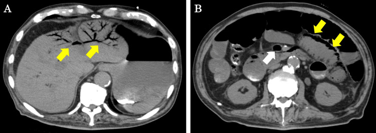
Figure 1. CT findings at initial examination.
(A) PVG can be observed mainly in the left lobe of the liver (yellow arrows). (B) Superior mesenteric venous gas (white arrow) and PI (yellow arrows) can be observed mainly in the proximal jejunum.
CT, computed tomography; PVG, portal venous gas; PI, pneumatosis intestinalis

