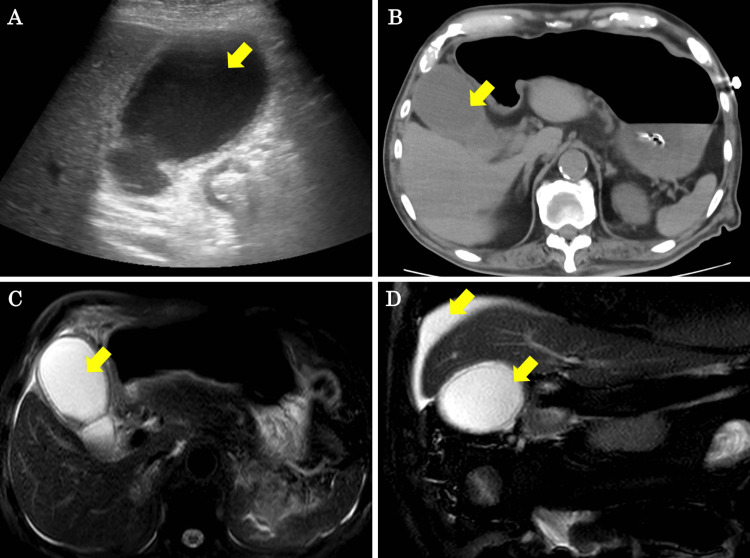
Figure 3. Abdominal ultrasound, CT, and MRCP findings on POD 6.
(A, B) Abdominal ultrasound and CT show gallbladder enlargement, thickening of the gallbladder wall, and gallbladder debris (yellow arrows). (C, D) MRCP shows fluid accumulation around the gallbladder and on the surface of the liver, as well as edema of the gallbladder wall (yellow arrows).
CT, computed tomography; POD, postoperative day; MRCP, magnetic resonance cholangiopancreatography

