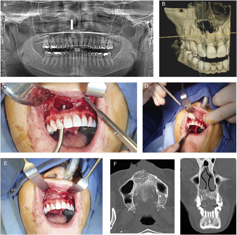
Figure 1.
Subject with anterior maxillary odontogenic cyst. (A) Preoperative panoramic radiograph, arrow points to lesion. (B) 3D reconstruction from cone beam computed tomography scan. (C) Intra operative photo after excision of lesion. (D) Placement of cellular bone matric allograft into defect. (E) Defect filled with cellular bone matrix. (F) CT scan taken 6 months after surgery. Left axial cuts and right coronal cuts. Note defect graft fill and consolidation.

