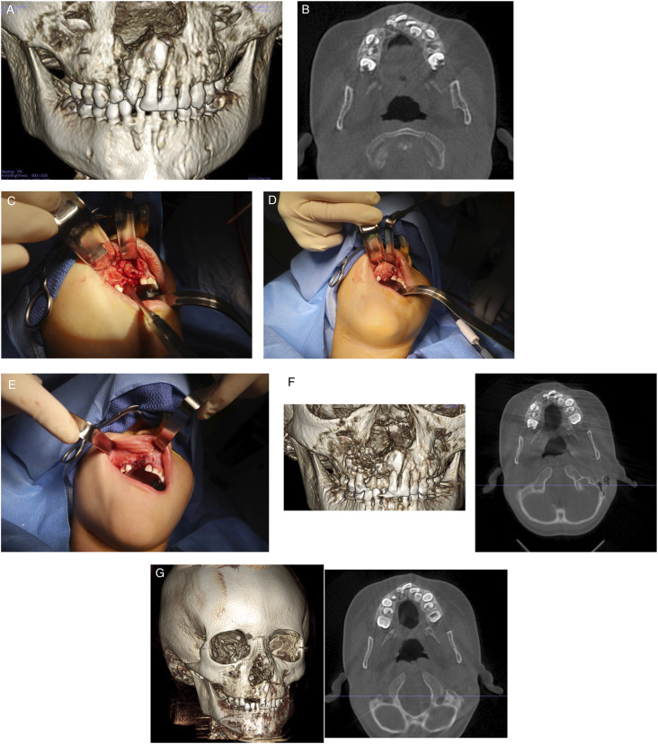
Figure 2.
Bone graft reconstruction of right cleft maxilla with cellular bone matrix and rh-BMP2. (A) Preoperative cone beam computed tomography scan demonstrating defect. (B) Axial scan demonstrating defect. (C) Intraoperative photograph demonstrating cleft maxilla after repair of residual oral nasal fistula. (D) Placement of cellular bone matrix and rh-BMP2 into cleft maxilla. (E) Tension free closure of oral mucosa. (F) Cone beam computed tomography scan one week after surgery, right 3D reconstruction, left axial cut. (G) Cone beam computed tomography scan taken 4 months after surgery, right 3D reconstruction, left axial cuts. Note union of the maxilla.

