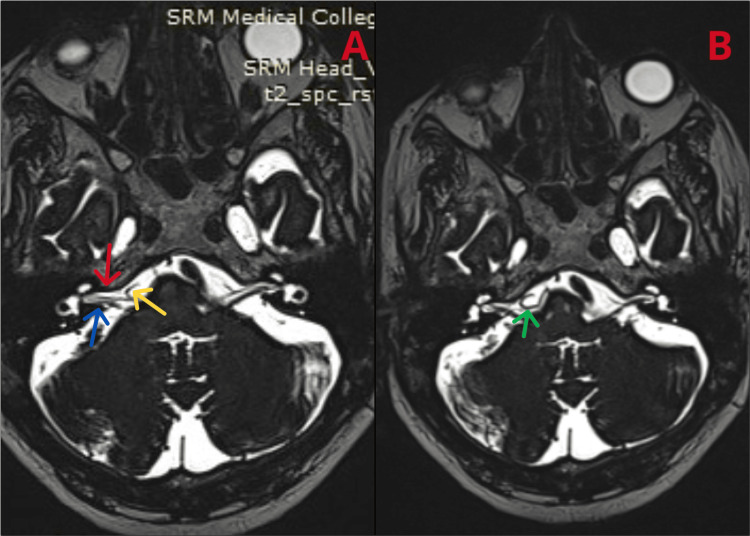
Figure 2. MRI of the brain revealing a (A) vascular loop (yellow arrow) of the AICA abutting the seventh (red arrow) and eighth (blue arrow) cranial nerve complexes on the right side in the CP angle cistern and (B) the AICA loop (green arrow).
MRI: magnetic resonance imaging; MRA: magnetic resonance angiography; MRV: magnetic resonance venography; AICA: anterior inferior cerebellar artery; CP: cerebellopontine

