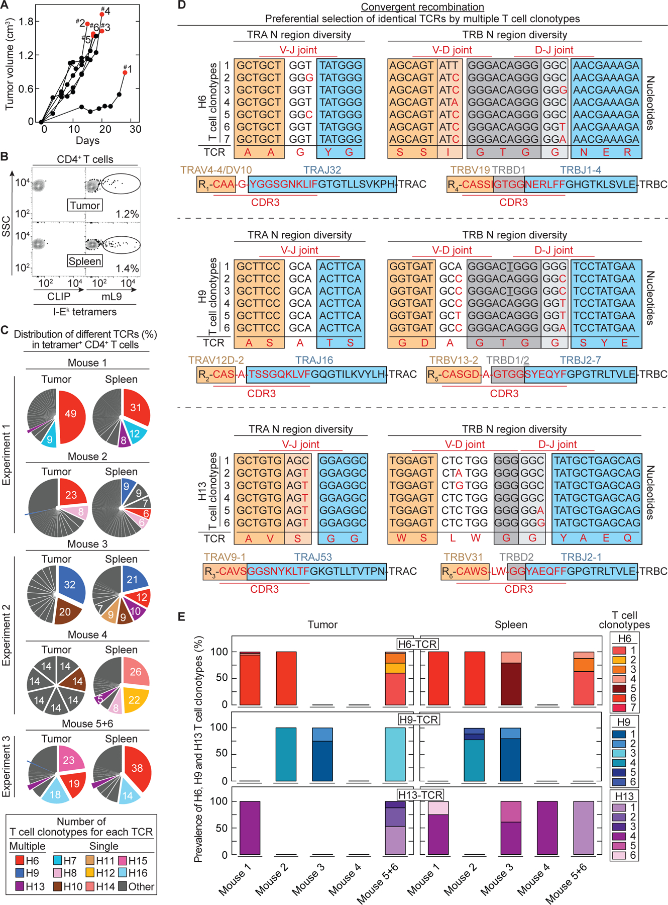
Fig. 1. Convergent recombination of different T cell clonotypes encoding identical, preferentially selected TCRs against the mutant neoantigen mL9.

(A) 6132A tumor fragments were injected s.c. into C3H/HeN mice. Six mice are shown which developed tumors after fragment injection (55%, 11/20 injected C3H/HeN mice) and were used for TCR analysis. Results were compiled from three independent experiments. Red dots indicate day of T cell analysis. (B) An example is shown of T cells isolated from spleen and tumor sorted for live, CD3+, CD4+ and mL9-tetramer+ specificity. Percentages of mL9-tetramer positive T cells are indicated. CLIP-tetramer staining was used as negative control. (C) Frequencies of paired TCR CDR3 amino acid sequences in mL9-teramer sorted CD4+ T cells obtained from tumor and spleen of the six analyzed mice. (D) Identification of different T cell clonotypes encoding an identical TCR based on N nucleotide sequence diversity in the TRA and TRB V(D)J joints. This was determined for the TCRs H6 (upper TCR), H9 (middle TCR) and H13 (bottom TCR). (E) Frequency of the different T cell clonotypes encoding an identical TCR (either H6, H9 or H13) among the analyzed mice.
