Fig. 1.
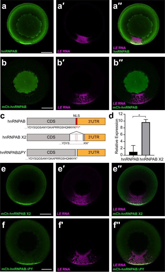
hnRNPAB X2 is a novel cytoplasmic splice isoform of hnRNPAB that is enriched in L-bodies. (a) Stage II oocytes were microinjected with Cy5-labeled LE RNA (magenta, a′) and immunostained for endogenous hnRNPAB (green, a) using antibodies raised against Xenopus hnRNPAB30. The overlap is shown in a′′. (b) Cy5-labeled LE RNA (magenta, b′) was microinjected into stage II oocytes expressing mCherry-tagged canonical hnRNPAB, as detected by immunostaining with anti-RFP (green, b). The overlap is shown in b′′. (c) Schematics of canonical hnRNPAB, hnRNPAB X2 and hnRNPABΔPY. C-terminal sequence is shown below each, with the PY NLS indicated in red, the protein coding sequence (CDS) in gray, and the 3′UTR shown in orange. (d) RNA isolated from stage II oocytes was used to measure the relative expression of hnRNPAB X2 compared to canonical hnRNPAB by qPCR. ΔCt values were calculated normalizing to refence gene vg1 (* indicates p < 0.05 by T-test). Error bars represent standard deviation of the mean, n = 3. (e) Cy5-labeled LE RNA (magenta, e′) was microinjected into stage II oocytes expressing mCherry-tagged hnRNPAB X2, as detected by immunostaining with anti-RFP (green, e). The overlap is shown in e′′. (f) Cy5-labeled LE RNA (magenta, e′) was microinjected into stage II oocytes expressing mCherry-tagged hnRNPABΔPY, as detected by immunostaining with anti-RFP (green, e). The overlap is shown in e′′. Confocal sections (a-b, e-f) are shown with the vegetal hemisphere at the bottom; scale bars = 100 μm.

