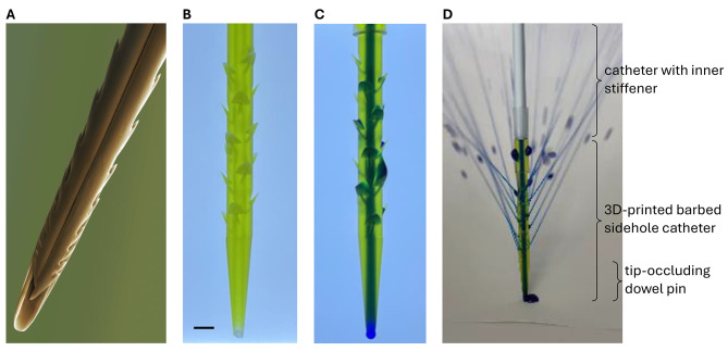
Fig. 3.
Barbed sidehole catheter. The barbs retain the catheter in the tumor, and the sideholes allow intratumoral drug infusion. A. Colorized SEM image of a honey bee stinger, showing the barbs that retain the stinger at the venom injection site (Image credit: Power & Syred / Science Photo Library). B. Backlit photograph of the translucent barbed sidehole catheter. Scale bar: 2 mm. C. Methylene blue was injected into the catheter, to show the main catheter channel, as well as the sidehole channels. D. Fluid jets emerging from the barbed sidehole catheter.

