
Figure 1.
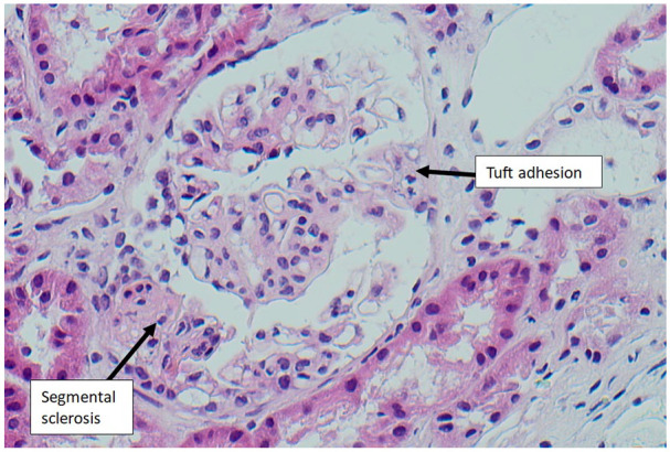
Light microscopy (haematoxylin and eosin stain, 200× magnification) of glomerulus showing features of FSGS-NOS, with segmental sclerotic lesion and adhesion to the overlying Bowman’s capsule (tuft adhesion).
FSGS-NOS, focal segmental glomerulosclerosis-not otherwise specified.