. 2024 Nov 4;5(12):1259–1270. doi: 10.1039/d4cb00174e

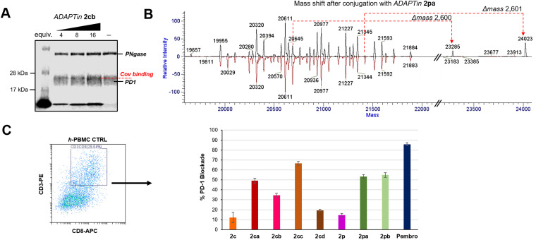
Fig. 5. Electrophilic ADAPTins activity on the PD1 Protein. (A) Denaturing SDS-PAGE gel analysis showing a dose-dependence conjugation of 2cb to PD1 after incubation (12 h, 37 °C). No conjugation observed with 2c (negative control). (B) Mass spectrometry characterization of PD1 conjugation with 2pa (conjugation reaction with protein : peptide 55 : 220 μM, pH 8.0, 48 h). Deconvolution of the PD1 glycol-forms before (bottom red) and after (top black) conjugation with 2pa. (C) In cellulo PD1 blockade by covalent ADAPTins2ca–cd and 2pa–pbversus non-covalent analogs 2c and 2p and the full antibody control pembrolizumab. Cell surface level of PD1 blockade in PBMC cultures (30 h) determined by flow-cytometry analysis. Cells were gated on CD45+ and CD3+/CD8+ cells were selected from the CD45+ fraction; error bars indicate the mean PD1%-blockade ± SD (n = 3).

Fig. 5