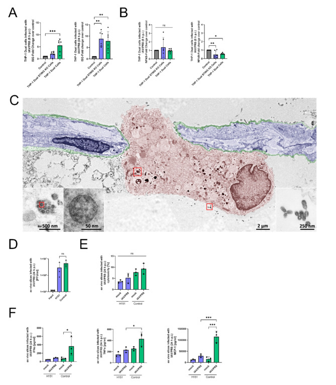
Fig. 3.
STING is necessary to induce interferon response in macrophages. (A) IFN stimulator gene (ISG) reporter fold change of THP-1 Dual and THP-1 Dual STING KO cells infected with IAV/PR8 at timepoints 8 h p.i. and 24 h p.i. The results of six biological replicates of each cell line is shown. Significance was calculated with One-way ANOVA (* p ≤ 0.05, ** p ≤ 0.01, *** p ≤ 0.001). (B) Transcriptional activity of NFκB pathway in THP-1 Dual and THP-1 Dual STING KO cells infected with IAV/PR8. Significance was calculated with One-way ANOVA (* p ≤ 0.05, ** p ≤ 0.01). (C) SEMpicture showing IAV on the surface and intracellular of an alveolar macrophage. Colors were added manually. (D) Viral titer of ex vivo lung slices infected with IAV/PR8 24 h p.i. was detected by standard plaque assay shown with logarithmic scaling. The results of three biological replicates are shown. Significance was calculated with One-way ANOVA (ns p ≥ 0.05). (E) LDH was measured in the supernatants of ex vivo slices and presented as percentage of the positive control. Significance was calculated with One-way ANOVA (ns p ≥ 0.05). (F) Cytokines IFNα, TNFα and MCP-1 were measured in the supernatants. Significance was calculated with One-way ANOVA (* p ≤ 0.05, ** p ≤ 0.01, *** p ≤ 0.001)

