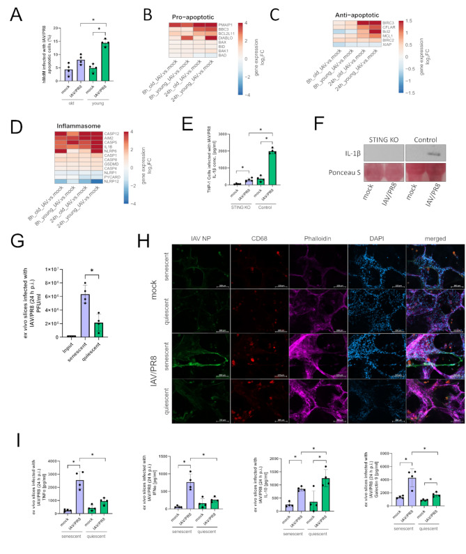
Fig. 5.
Apoptosis and inflammasome signaling pathways are altered in old macrophages. (A) Apoptotic cells were quantified via flow-cytometry. The results of four biological replicates in each group are shown. Significance was calculated with the Mann-Whitney U test (* p ≤ 0.05). (B) Heatmaps showing proapoptotic, (C) antiapoptotic genes (D) and genes of inflammasome signaling. Gene expression is presented over log2 fold change. At least one DEG per gene per condition had a significant log2 fold change. (E) Concentration of IL-1β was measured in the supernatants of infected THP-1 Dual and THP-1 Dual STING KO cells 24 h p.i. Results were confirmed by western blotting using the indicated antibodies. Significance was calculated with the Mann-Whitney U test (* p ≤ 0.05). (F) Supernatants of THP-1 cells were analyzed by western blot using the indicated antibody and staining. The experiment was repeated 4 times and blot shown as representative. (G) Viral titer of ex vivo lung slices was determined by standard plaque assay. Significance was calculated with the Mann-Whitney U test (* p ≤ 0.05). (H) Immunofluorescence staining of infected ex vivo lung slices stained with antibodies against IAV NP, CD68, phalloidin and DAPI. (I) Cytokines in the supernatants of senescent and quiescent ex vivo lung slices were measured. Significance was calculated with the Mann-Whitney U test (* p ≤ 0.05)

