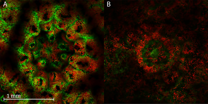
Figure 2. Confocal images of a live and dead coral fragment.
Confocal images of a polyp from a live and dead coral fragment after 24 h in culture. (A) Live polyp, 24 h: Merged image of the autofluorescent symbiotic algae (red) and the autofluorescent green fluorescent protein of the coral (GFP, green). Note how tightly organized the GFP and symbionts are around the polyp mouth and tentacles. The confocal image clearly shows the morphology of the polyp skeleton and tentacles. (B) Dead polyp, 24 h: Merged image of the autofluorescent symbiotic algae (red) and the autofluorescent GFP of the coral (green). Note the disorganized pattern of the GFP and symbionts fluorescence around the polyp and tentacles. The polyp skeleton and tentacles were degraded and appear blurred. The symbiont fluorescence is scattered across the image and the GFP is blurred.

