. 2024 Nov 7;24(24):5360–5373. doi: 10.1039/d4lc00659c

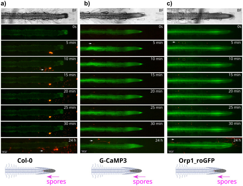
Fig. 6. P. capsici zoospore localisation patterns in wild type Col-0 and transgenic G-CaMP3 and Orp1_roGFP A. thaliana roots. Pathogen spores were passively injected through the tip-site of the bi-dfRC (inlet C), over 24 h. Bright field (BF) channel displayed above. Schematic diagrams depicting treatment application (white arrow) displayed below. Key events including zoospore localisation, germination and protrusion into root tissue are highlighted with magenta arrows. Original video files are 0.2 fps. (a) Zoospore release from a sporangium situated below a Col-0 primary root, within the bi-dfRC microchannel. Zoospores localise to the root tip and maturation/elongation (ME) zone over 30 min. Infection is observed at the elongation/differentiation zone (ED) of the primary root after 24 h. No root signalling was observed. (b) Zoospore localisation to a G-CaMP3 root hair within 5 min. A strong Ca2+ burst indicated by the G-CaMP3 was observed within 5 min following active injection of zoospores, persisting for 30 min. Infection at the ED zone was observed 24 hours following exposure. Whole root Ca2+ levels remain elevated 24 h following infection. (c) Zoospores localise to Orp1_roGFP root hairs, ME and ED zones within 5 min. A gradual increase in H2O2 was observed over 30 min, following the passive injection of pathogen spores at the tip site (inlet C) of the bi-dfRC. Infection is shown at the tip and DZ of the primary root after 24 h. Whole root ROS upregulation H2O2 was observed 24 h following infection.

Fig. 6