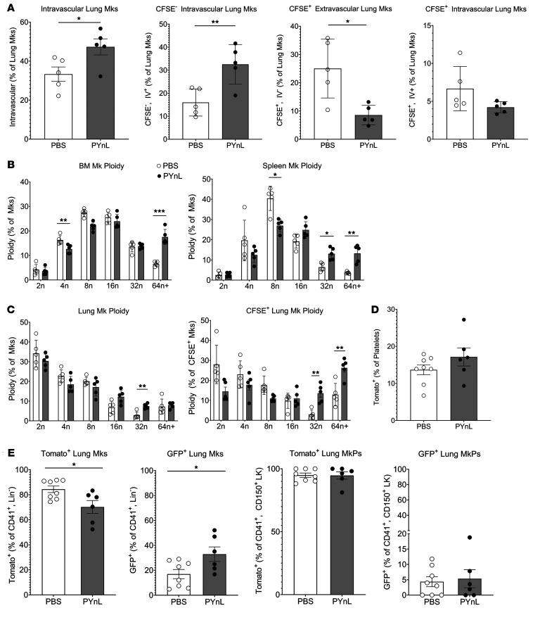
Figure 6. Mks migrate to the lung to respond to increased platelet demand.
(A) Mice were given CFSE o.p. and infected with PYnL, and on day 14, total Mks and CFSE+ and CFSE– intravascular (CD42d i.v. -positive) and extravascular (CD42d i.v. –negative) Mks were quantified. The increase in total intravascular Mks was driven by CFSE– extrapulmonary Mks. On day 14, there was a decrease in CFSE+ extra- and intravascular Mks (n = 5 per group, representative shown from 2 independent experiments). (B and C) With PYnL infection, there was an increase in higher ploidy CFSE+ Mks in (B) BM and spleen as well as in (C) lung, including CFSE+ Mks (n = 5 per group, representative shown from 2 independent experiments). (D) FlkSwitch mice infected with PYnL had no significant change in Tomato+Flt3– platelets on day 14 after infection, but (E) the percentage of lung Mks that were Tomato+Flt3– slightly declined and that of GFP+Flt3+ Mks increased slightly compared with uninfected controls. There was no change in MkPs, indicating an influx of mature Mks from the BM that was largely Tomato+Flt3– (n = 6–8 per group from 2 independent experiments). Data indicate the mean ± SEM. *P < 0.05, **P < 0.01, and ***P < 0.001, by unpaired, 2-tailed t test (A, D, and E) and multiple t tests with Holm-Šidák correction (B and C).

