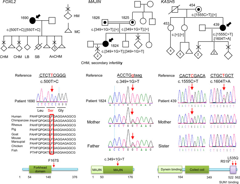
Figure 1. Pedigrees, Sanger sequencing, and segregation of the variants in FOXL2, MAJIN, and KASH5.
The probands are indicated by arrows. Amino acid numbering is given below the protein structure. On the protein structure, the red arrows indicate the positions of the variants seen in a recessive state. HM, hydatidiform mole; CHM, complete HM; AnCHM, androgenetic CHM; LB, live birth; SB, stillbirth; MC, miscarriage.

