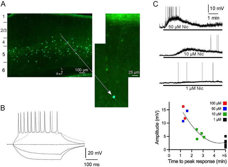
Figure 1.
α2 nAChRs-expressing neurons in A1: distribution, intrinsic electrophysiology and response to nicotine, in vitro. (A) Neurons with EGFP are nonpyramidal, primarily in layer 5, and have processes that are largely restricted to layer 5, or extend vertically towards layer 1 (see higher power view) where they terminate in a dense band of fluorescent fibers. The imaged section is cut along the thalamocortical (near horizontal) plane and numbers on the left indicate approximate boundaries of layers (see Methods); arrows indicate lateral and rostral directions. (B) In vitro whole-cell recording showing membrane responses of a fluorescent neuron to intracellular current pulses. (C) In vitro response to bath applied nicotine (1–100 μM for >5 min) for individual neurons (top; action potentials truncated) and in group dose–response data (bottom).

