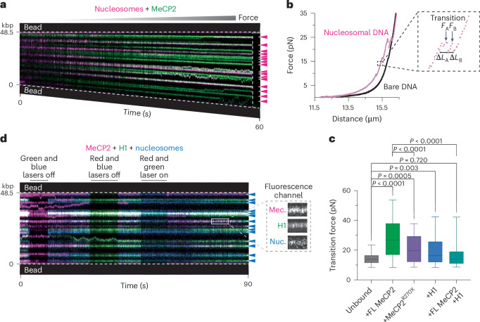
Fig. 5. MeCP2 enhances the mechanical stability of nucleosomes.
a, Representative kymograph of an LD655-nucleosome-containing unmethylated DNA tether bound with Cy3-MeCP2 and pulled to high forces by gradually increasing the inter-bead distance. The vertical dotted line denotes the time when the tether ruptured. The arrowheads denote nucleosome positions. b, Representative force–distance curve of a MeCP2-bound nucleosome-containing DNA tether (red) showing force-induced transitions overlaid on a force–distance curve of a MeCP2-bound bare DNA tether (black). The inset shows a zoom-in view of two example transitions for which the distance change (ΔL) and the transition force are recorded. The experiment was independently repeated with 7 (nucleosomal DNA) and 5 (bare DNA) tethers yielding similar results. c, Distribution of transition forces recorded from force–distance curves of nucleosomal DNA tethers with no MeCP2 or H1 bound (n = 84 from 5 independent tethers), bound with FL MeCP2 (n = 107 from 7 independent tethers), MeCP2R270X (n = 68 from 7 independent tethers), H1 (n = 106 from 10 independent tethers) or both FL MeCP2 and H1 (n = 81 from 8 independent tethers). The box boundaries represent the 25th to 75th percentiles, the middle bar represents the median and the whiskers represent the minimum and maximum values. Significance was calculated using a one-way ANOVA with Tukey’s test for multiple comparisons. d, Representative kymograph of an unmethylated DNA tether containing AF488-labeled nucleosomes and incubated with Cy5-labeled FL MeCP2 and Cy3-labeled H1. Lasers were switched on and off to confirm fluorescence signals from each channel. The arrowheads denote nucleosome positions. The inset shows a zoom-in view of individual fluorescence channels at a nucleosome site (Nuc.) where MeCP2 (Mec.) and H1 colocalized.

