Fig. 1.
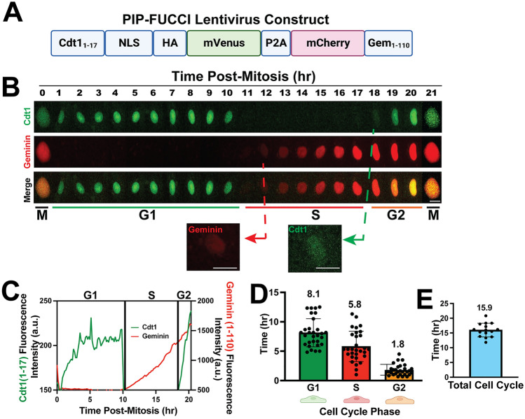
PIP-FUCCI Lentivirus reports cell cycle status in HUVEC. A PIP-FUCCI lentivirus construct. NLS, nuclear localization signal; HA, HA tag; P2A, self-cleaving peptide P2A; Gem, geminin. B Representative time-lapse images of PIP-FUCCI transduced HUVEC, showing mVenus-Cdt11-17 (green) and mCherry-Geminin1-110 (red) expression hourly from end of cytokinesis (M) through next cytokinesis. Scale bar, 20 μm. C Quantification of PIP-FUCCI fluorescence intensity/time from cell in B. D Average time spent in each cell cycle phase (hr). (n = 30 cells per phase from 3 replicate movies) E. Total endothelial cell cycle length, time between mitoses measured by H2B-CFP. (n = 15 cells from 3 replicate movies)

