Fig. 1.
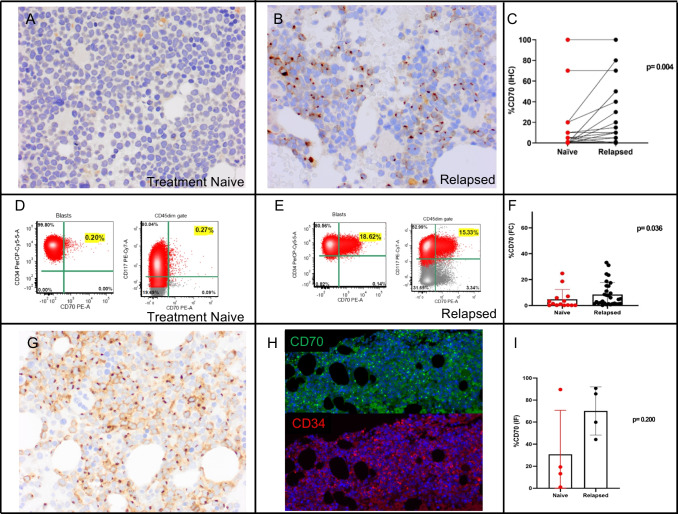
Examples of CD70 expression as detected by immunohistochemistry (IHC) and flow cytometry (FC) in treatment naïve and relapsed AML patients. A, B In this case, IHC for CD70 in a bone marrow clot was negative at diagnosis (A) and detected in about 50% of the blasts at the relapse one year later (B); a paired analysis of naïve and relapsed samples shows a significantly higher expression in the relapsed group (p = 0.004) (C). D, E FC of a naïve treated sample (D) shows expression of CD70 in 0.20% of the CD34+ blasts and 0.27% of CD117+ blasts, while at time of the relapsed demonstrates positivity for CD70 in 18.6% of the CD34+ blast population and in 15.3% of the CD117+ (E); an analysis using FC to compare naïve and relapsed samples shows a statistically significant higher expression in relapsed samples (p = 0.036) (F). G, H IHC for CD70 in a bone marrow clot of a newly diagnosed AML shows positivity in most of the blasts (G) and the dual immunofluorescence (IF) for CD34 (red)/CD70 (green) in the same specimen shows positivity in 89.6% of the blasts as quantified by digital imaging (H). A comparison using dual IF showed a higher expression in relapsed samples in comparison with naïve treated, although not statistically significant, likely due to the low number of samples with double IHC and IF staining (I). Note that the immunostaining pattern includes staining of the cytoplasmic membrane and a Golgi/paranuclear stain in most of positive blasts. Blasts with only a Golgi stain pattern are also seen, and if these blasts express CD70 at the level of the cellular membrane is unknown currently

