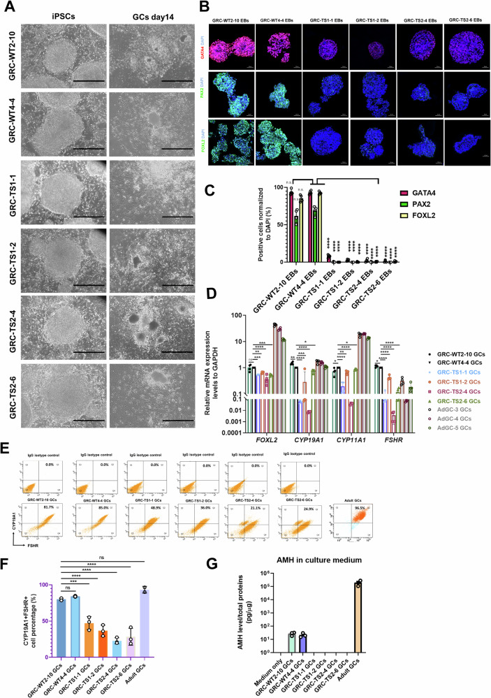
Fig. 2. Granulosa cells derived from Turner syndrome patients display differentiation defects and downregulate anti-Müllerian hormone (AMH) secretion capabilities.
A Representative brightfield images of hiPSCs and the differentiated GCs derived from healthy control (WT) fibroblasts and Turner syndrome (TS) fibroblasts. Scale bar, 1000 μm. The experimental GC differentiation were performed three times independently. B Representative immunofluorescence (IF) staining characterizations of GATA4 (mesoderm marker), PAX2 (intermediate mesoderm marker), and FOXL2 (GC-specific marker) in WT-GC and TS-GC clones during each time point of GC differentiation. Scale bar, 50 μm. The experimental GC differentiation were performed at least three times independently. C Quantification of fluorescence images of (B). Bars indicate the mean ± SD (n = 4). ****p < 0.0001 vs. GRC-WT4-4 EBs. D Quantitative analysis of the expression of specific genes in GCs differentiated from iPSCs (day 14) and in luteinized cumulus granulosa cells. Bars indicate the mean ± SD (n = 4). *p < 0.05, **p < 0.01, ***p < 0.001, and ****p < 0.0001 vs. GRC-WT4-4 GCs. E Flow cytometry analysis of GC markers, CYP19A1 and FSHR between healthy control (WT) GCs and TS-GCs. Bars indicate the mean ± SD (n = 3). The luteinized cumulus granulosa cells from two adult donors served as staining positive control samples. F The flow cytometry quantification result of (E). Bars indicate the mean ± SD (n = 3 for iPSC-derived GCs; n = 2 for adult GCs). ***p < 0.001, and ****p < 0.0001 vs. GRC-WT2-10 GCs. G AMH ELISA assays were performed to quantify the AMH levels in the culture supernatants collected from healthy control (WT) GCs and TS-GCs cultured for 3 days. The supernatant collected from the luteinized cumulus granulosa cells served as positive control cells. Secreted AMH levels were normalized to the total protein amounts of the cultured cells. Bars indicate the mean ± SD (n = 3).

