
Figure 3.
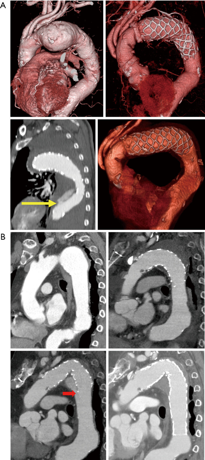
CT image of patients with aortic events. (A) CT angiography image of a patient with dissection. (B) CT angiography image of a patient with type Ib endoleak. The yellow arrow shows the distal end of the Frozenix J-graft, and the red arrow shows a type Ib endoleak. Figure 3A are reused with permission from AME Publishing Company (12). CT, computed tomography.