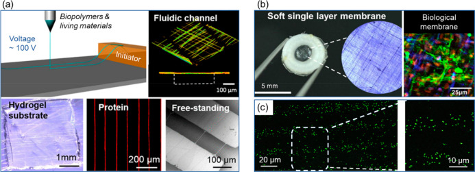
Figure 4.
Low voltage electrospinning of biopolymers and living materials. (a) A schematic illustration of the LEP process and microscope images showing various fiber materials and patterns. Adapted and reprinted with permission from ref (36). Copyright 2016 American Chemical Society. (b) A suspended ECM-laden fiber membrane patterned on a 3D printed frame and immunofluorescence image of cell coculture of glomerular endothelial cells and podocytes on the membrane (red fluorescence: nuclei; green fluorescence: VE-Cad; blue fluorescence: podocytes). Adapted with permission from ref (46). Copyright 2018 Elsevier. (c) Confocal images of LEP patterned E. coli fiber arrays on glass. Reprinted with permission from ref (36). Copyright 2016 American Chemical Society.

