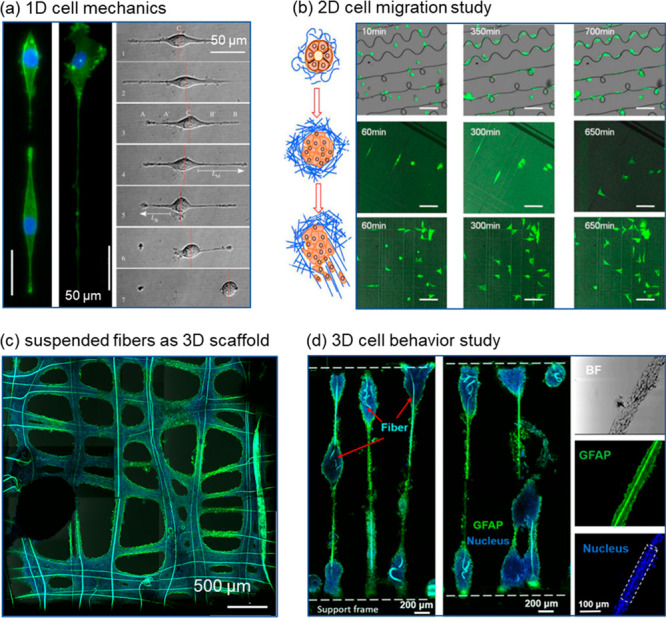
Figure 6.

1D to 3D culture to study cell mechanics. (a) Time lapse images of the EA.hy926 endothelial cell migration process on 1D gelatin fiber (green fluorescence: F-actin; blue fluorescence: nuclei). Reprinted with permission from ref (51). Copyright 2014 The Royal Society. (b) Time lapse images of GFP-tagged MDA-MB-231 cancer cells migrating on 2D polystyrene fiber networks of various patterns. (c) Human glioblastoma cells U87 aggregated on gelatin fibers (green fluorescence: GFAP, a glial cytoskeletal marker; blue fluorescence: nuclei). Reprinted with permission from ref (37). Copyright 2019 American Chemical Society. (d) Immunofluorescence images of an ellipsoid-on-string formed by human glioblastoma U87 cells along the suspended gelatin microfibers (green fluorescence: GFAP, a glial cytoskeletal marker; blue fluorescence: nuclei). Reprinted with permission from ref (15). Copyright 2021 IOP Science.
