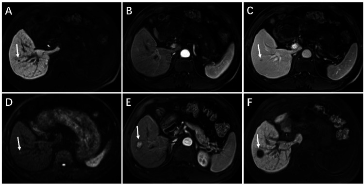
Figure 2.
Hypervascular transformation of a hepatobiliary phase hypointense nodule without arterial phase hyperenhancement manifested with the combination of DWI hyperintensity and PVP hypointensity in a 64-year-old man with hepatitis B–induced liver cirrhosis. On initial gadoxetic acid-enhance MRI (A-D), a 5-mm nodule shows hypointensity on 20-min hepatobiliary phase imaging (A, arrow), isointensity on arterial phase T1-weighted imaging (B), hypointensity on PVP imaging (C, arrow), and hyperintensity on DWI (b = 500 s/mm2) (D, arrow). Follow-up gadoxetic acid-enhanced MRI obtained 445 days later, the nodule shows marked enlargement as measuring 18-mm, unequivocally hyperenhancement than surrounding liver on arterial phase imaging (E, arrow), and hypointensity on 20-min hepatobiliary phase imaging (F, arrow). Nodule specimens after surgical resection confirmed the diagnosis of HCC. Abbreviations: DWI: diffusion-weighted imaging; PVP: portal venous phase; HCC: hepatocellular carcinoma.

