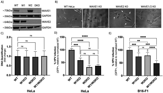
Fig 2. WAVE1 knockout (W1KO), W2KO, and W1/W2KO alters cellular morphology, but not proliferation, and inhibits HPV16 infection in multiple cell lines.
(A) WAVE1 (W1) WAVE2 (W2) or both (DKO) proteins were knocked out in wild type (WT) HeLa cells via CRISPR/Cas9 and confirmed by Western blotting. (B) Representative phase-contrast images of WT, W1KO, W2KO, and W1/W2KO HeLa cells were taken on the FloID Cell Imaging Station (20x magnification, scale bar = 50μm). (C) W1KO, W2KO, and W1/W2KO HeLa cells were seeded in equal amounts, grown for 48 hours, and then analyzed for differences in DNA quantity via CyQUANT Cell Proliferation Assay (Thermo Fisher) compared to WT. (D and E) WT, W1KO, W2KO, and W1/W2KO HeLa or B16-F1 cells were treated with HPV16 PsVs (TCID30) containing a GFP reporter plasmid. The percentage of infected cells (based on GFP reporter gene expression) was measured at 48 hours post infection via flow cytometry. Background from mock infected cells was subtracted. For HeLa cells, at least 2 independent clones of each knockout were screened for consistent inhibition of HPV16 infection. Each bar represents three biological repeats comprised of technical triplicates and show DNA quantification over 48 hours (Panel C) or the mean %GFP+ cells ± standard deviation (n=3, normalized to WT) (Panels D and E). 1-way ANOVA with Dunnett’s multiple comparisons test was used to statistically determine significance (ns=not significant, **p<0.001, ***p<0.0001, ****p<0.0001).

