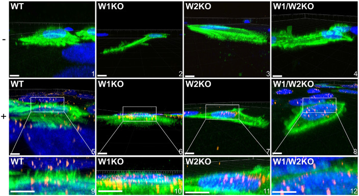
Fig 7. Knockout of WAVE1, WAVE2, or both, prevents HPV16 stimulated HeLa cells from expressing dorsal surface actin protrusions.
Cells were prepared as described in Fig 5 however, cells were not permeabilized during immunostaining. Either untreated (top row, − symbol) or HPV16 infected WT, W1KO, or W1/W2KO HeLa cells (10 ng/1E6 cells) (middle row, + symbol) treated with CellLight Actin-GFP were imaged via laser scanning confocal microscopy to obtain Z-stacks. Z-stacks were then stitched together and rotated to view the XZ oriented volume. Scale: images 1, 2, 4-7 = 10 μm; image 3 = 8 μm; image 8 = 14 μm. 22 cells were analyzed per condition.

