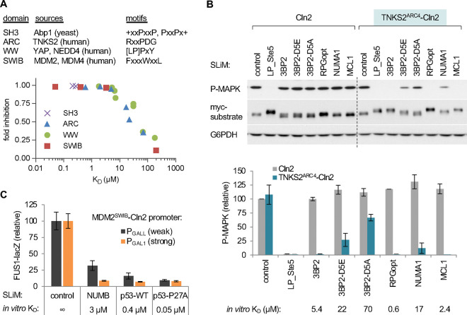
Figure 2. Test cases and low-throughput assays of SLiM binding via the SIMBA system.
(A) Top, tested domains and their motif types. Bottom, summary of results presented in Figures 2B and S1D–F. The plot shows functional strength measured by SIMBA in vivo vs. previously measured affinities in vitro. Error bars are omitted for clarity. See also Figure S1C.
(B) Cln2 and a TNKS2ARC4-Cln2 fusion were coexpressed with Ste20Ste5PM derivatives harboring known TNKS2ARC4-binding peptides, and then pheromone signaling was assayed. Top, representive blots; SLiM binding blocks activation of the MAPK Fus3 (P-MAPK) and promotes a mobility shift of the myc-tagged Ste20Ste5PM substrate. Bottom, quantification of results (mean ± SD; n = 2 [Cln2] or n = 6 [TNKS2ARC4-Cln2]), compared with in vitro binding affinity [54]. See also Figure S1A–B.
(C) Improved resolution of strong interactions by expressing the MDM2SWIB domain from a weaker promoter (PGALL) vs. the full strength promoter (PGAL1). Bars, mean ± SD (n = 4). See also Figure S1C.

