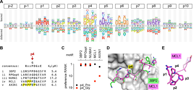
Figure 4. Local context affects residue preferences at some positions.
(A) Logo comparing the residue preferences at each peptide position in the context of 5 different parent peptides that bind TNKS2ARC4 (labeled i-v; see panel [B]).
(B) Sequence and KD of parent peptides (i-v) analyzed in panel (A). Yellow, Pro residues flanking p4 in MCL1 and AXIN1. Blue, residues that are atypically preferred in MCL1 and AXIN1.
(C) Preference ranks of Pro and Gly at p4 in 5 different parent motifs.
(D) Similar trajectories and p4 contacts of 3BP2 and MCL1 peptides bound to TNKS2ARC4 [54]. PDB IDs: 3twr, 3twu.
(E) Rotated view of the TNKS2ARC4-bound MCL1 peptide, showing left-handed trajectory of poly-proline sequence from p2 to p4

