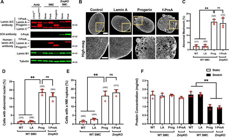
Fig. 2. Progerin and farnesyl-prelamin A have similar effects in cultured SMCs.
A. Western blot comparing the expression of nuclear lamins in the mouse aorta and in mouse SMCs expressing human lamin A, human progerin, and human farnesyl-prelamin A. Nuclear lamins were detected with antibodies that bind to mouse and human lamin A/C, farnesyl-prelamin A (clone 3C8), human lamin A/C, and lamin B1. Tubulin was measured as a loading control. B. Representative high-resolution confocal microscopy images showing the protein meshworks formed by human versions of lamin A, progerin, and farnesyl-prelamin A (f-PreA). Scale bar, 5 μm. The boxed regions are shown at higher magnification below. Scale bar, 2 μm. C. Nuclei with an abnormal meshwork in SMCs expressing human lamin A (LA), progerin (Prog), and farnesyl-prelamin A (f-PreA). Mean ± SEM (n = 3 experiments). ANOVA. **, P < 0.01. ns, not significant. D. Abnormal nuclear shape in SMCs expressing human versions of lamin A, progerin, and farnesyl-prelamin A. Mean ± SEM (n = 3 experiments). ANOVA. **, P < 0.01. ns, not significant. E. Nuclear membrane (NM) ruptures in SMCs expressing human versions of lamin A, progerin, and farnesyl-prelamin A. Mean ± SEM (n = 3 experiments). ANOVA. **, P < 0.01. ns, not significant. F. Cell death in SMCs expressing human versions of lamin A, progerin, or farnesyl-prelamin A. SMCs were cultured on PDMS membranes and exposed to static (open bars) or cyclical stretching conditions (closed bars) for 24 h. The fraction of cells remaining on the membranes were quantified by measuring protein concentration. Mean ± SEM (n = 3 experiments). ANOVA. **, P < 0.01. For the data reported in C–E, the number of nuclei or cells examined are shown in parentheses.

