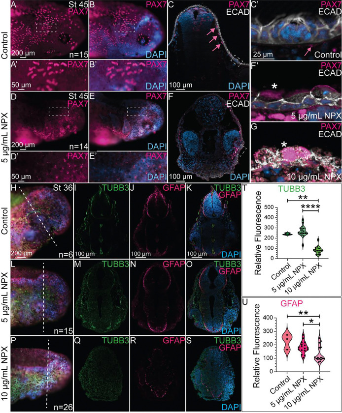
Figure 6. NPX alters PAX7 expression and the formation of ectodermally-derived structures..
IHC for PAX7 and ECAD in (A, B, D, and E) whole mount and (C, F) transverse sections. (A’, B’, D’, and E’) Zoom in from A, B, D, and E (dashed boxes). (C) In control embryos, PAX7 and ECAD are localized to pigment cells and superficial sensory organs. Pink arrows highlight subepidermal localization of PAX7+ pigment cells. Dashed box identifies an ECAD+ sensory organ. (C’, F’) Zoom in from sections in C and F (dashed boxes). The pink arrows in panel C’ again highlights the position of PAX7 expression relative to the sensory structures in the skin. (F’, G) Sensory organs from exposed embryos develop abnormally by stage 45. Asterisks indicate mislocalization of PAX7 expression. IHC for TUBB3 and GFAP shown in (H, L, P) wholemount and (I-K, M-O, and Q-S) transverse sections in (H-K) control embryos and (L-S) NPX exposed embryos. (H, L, P) Dashed lines show the axial level of transverse sections. (I-K, M-O, and Q-S) With exposure to increasing concentrations of NPX, there is corresponding reductions in expression of TUBB3 (neurons) and GFAP (astrocytes/glia), in the neural tube. (T, U) Quantification of TUBB3 and GFAP expression in cross-section. The sample sizes used for quantification of both TUBB3 and GFAP expression were n=1, n=2, and n=2 embryos for the control, 5 μg/mL, and 10 μg/mL NPX groups. An average of 7 transverse sections per embryo were quantified for either TUBB3 or GFAP expression.

