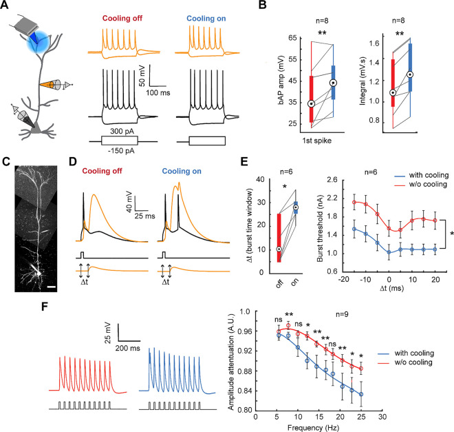
Fig. 4. Somato-dendritic coupling modulation as a function of temperature.
A) Schematic of the dual somatic and apical dendritic patch clamp recording while cooling the tuft dendrite (left). Prototypical spike trains recorded at the soma (bottom) and apical dendrite (top) under moderate focal cooling of the tuft dendrite at T=30°C (right) and T=35°C (left).
B) The amplitude comparison of the first bAP spike (left) (Wilcoxon signed rank test, n=8 neurons, p=0.0078), and the integral (right) (Wilcoxon signed rank test, n=8 neurons, p=0.0039) comparing conditions with and without mild focal cooling.
C) Exemplar layer 5 pyramidal neuron loaded with Alexa 488 (100 μM) under somato-dendritic patch clamping (scale bars, 50 μm).
D) The combination of current injection at the soma and the apical dendrites EPSP- like depolarization separated by a time interval (Δt) of 10 ms evoked a burst following the onset of Ca2+ -AP in the apical dendrite while the tuft dendrite is focally cooled.
E) The time duration (Δt) (left) and threshold needed to generate the burst (right) significantly increased and decreased, respectively, under mild focal cooling. (Wilcoxon signed rank test, n=6 neurons, *p<0.05). Error bars represent the standard error of the mean (SEM).
F) The amplitude attenuation at the dendrite (ratio of the amplitude of the last spike to the third spike in a train of bAPs generated by 0.8 nA pulses at the soma) with and without mild focal cooling (Wilcoxon signed rank test, n=9 neurons for all points except for 10 Hz and 12.2 Hz where n=7, *p<0.05, **p<0.01). Error bars represent the standard error of the mean (SEM).

