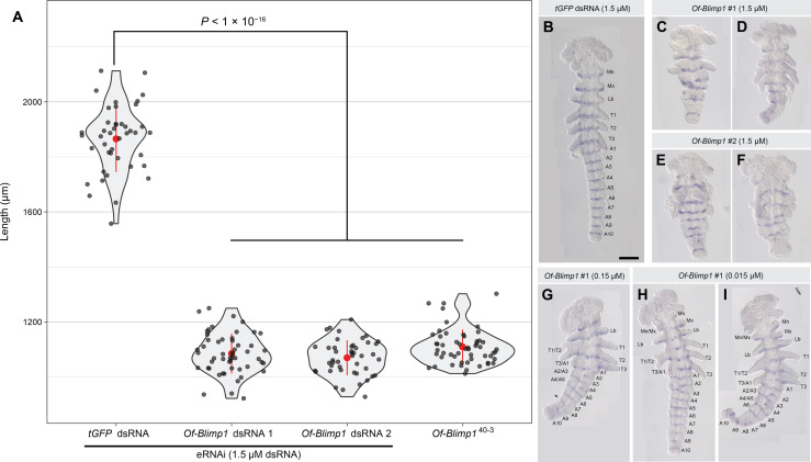
Fig. 3. Knockdown of Of-Blimp1 results in shortened embryos and segmental fusions suggesting PR-like function.
(A) Violin plots displaying the distribution of embryonic lengths in control (tGFP dsRNA-injected), Of-Blimp1 eRNAi embryos (67 to 71.5 hours AEL), and Of-Blimp140–3 presumptive homozygotes. The means and SD are shown in red. The data are plotted in gray dots. (B) tGFP dsRNA–injected embryo with one inv stripe in each of the six appendage bearing segments and each of the 10 abdominal segments. (C and D) Embryos injected with 1.5 μM Of-Blimp1 dsRNA-1 and (E and F) embryos injected with 1.5 μM Of-Blimp1 dsRNA-2 displaying reduced segment number and concomitant reduced inv stripe number. (G) Embryo injected with 0.15 μM Of-Blimp1 dsRNA-1. The right half of the embryo is mostly wild type, while segments are fused in the left half. Arrowhead marks a possible fusion of A7 and A8. (H and I) Embryos injected with 0.015 μM Of-Blimp1 dsRNA-1 displaying partial segment loss. The right half of both embryos exhibits wild-type segment number, while segment fusions are seen on the left side. All eRNAi embryos fixed at 67 to 71.5 hours AEL; Of-Blimp140-3 embryos fixed at 48-72 h AEL. Scale bar, (B) 200 μm.

