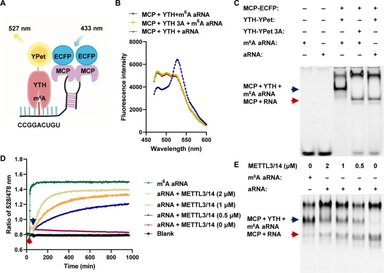
Fig. 1. In vitro FRET system for detecting m6A modifications on aRNAs.
(A) Schematic diagram of the FRET system based on m6A aRNAs. (B) Fluorescence spectrum after MCP-ECFP, YTH-YPet, or YTH-YPet-3A was mixed with aRNA or m6A aRNA. (C) Gel red staining of native polyacrylamide gel electrophoresis (PAGE) after MCP-ECFP, YTH-YPet, or YTH-YPet-3A was mixed with aRNA or m6A aRNA; (i) m6A aRNA, (ii) aRNA (iii) MCP-ECFP + YTH-YPet + m6A aRNA; (iv) MCP-ECFP + YTH-YPet-3A + m6A aRNA, and (v) MCP-ECFP + YTH-YPet + aRNA; blue arrow: the complex of YTH, MCP, and m6A aRNA; red arrow: the complex of MCP and RNA. (D) Ratio of 528/478-nm emission after MCP-ECFP and YTH-YPet were mixed; the red arrow indicates the addition of aRNA or m6A aRNA, and the blue arrow indicates the addition of METTL3/14 at different concentrations; n = 3. (E) Gel red staining of native PAGE for (D) (i) m6A aRNA + YTH-YPet + MCP-ECFP, (ii) aRNA + YTH-YPet + MCP-ECFP + METTL3 (2 μM), (iii) aRNA + YTH-YPet + MCP-ECFP + METTL3 (1 μM), (iv) aRNA + YTH-YPet + MCP-ECFP + METTL3 (0.5 μM), (v) aRNA + YTH-YPet + MCP-ECFP + METTL3 (0 μM); the blue arrow indicates the complex of YTH, MCP, and m6A aRNA; the red arrow indicates the complex of MCP and RNA.

