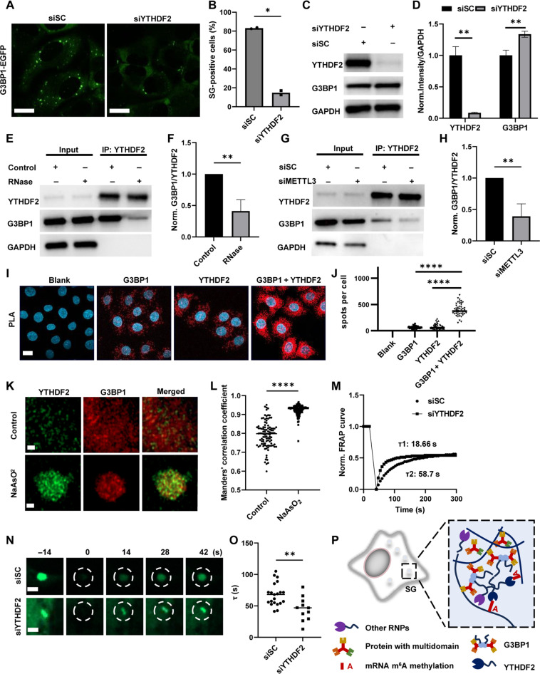
Fig. 7. YTHDF2 regulates the stability of SGs by interacting with G3BP1.
EGFP images (A) and percentage of SG-positive cells (B) in U2OS G3BP1-EGFP after siRNA treatment for 48 hours and NaAsO2 incubation for 1 hour. Scale bars, 15 μm. Cells with visible puncta labeled by EGFP in cytosol were counted as SG-positive cells. Unpaired t test with Welch’s correction (n = 2, P = 0.0136). (C) Protein levels of G3BP1, YTHDF2, and GAPDH in HeLa cells with or without siRNA treatment were determined via Western blot (WB). (D) Normalized YTHDF2/GAPDH and G3BP1/GAPDH intensities in (C). n = 3, unpaired t test with Welch’s correction (YTHDF2: P = 0.0073 and G3BP1: P = 0.0079). (E) Protein levels of G3BP1, YTHDF2, and GAPDH determined by WB after YTHDF2 pulling down in HeLa cells with or without RNase treatment. (F) Normalized G3BP1/YTHDF2 intensity in (E). Unpaired t test with Welch’s correction (n = 3, P = 0.0071). (G) Protein levels of G3BP1, YTHDF2, and GAPDH determined by WB after YTHDF2 pulling down in HeLa cells treated with siSC or siMETTL3. (H) Normalized G3BP1/YTHDF2 intensity in (G). Unpaired t test with Welch’s correction (n = 4, P = 0.0087). (I) PLA assays in HeLa cells incubated with/without G3BP1/YTHDF2 primary antibody. Scale bar, 10 μm. (J) Number of spots per cell in PLA assays. Unpaired t test with Welch’s correction (n = 50, P < 0.0001). (K) Expansion microscopy of YTHDF2 and G3BP1 with/without NaAsO2 treatment for 1 hour. Scale bars, 1 μm. (L) Manders’ values for interaction between G3BP1 and YTHDF2 in (K). Unpaired t test with Welch’s correction (n = 101, P < 0.0001). (M) Normalized FRAP curves of G3BP1-EGFP (dashed circles) in G3BP1-EGFP U2OS after siRNA treatment during FRAP. (N) G3BP1-EGFP images in (M). Scale bar, 1 μm. (O) τ of FRAP in (M). Unpaired t test with Welch’s correction (siSC: n = 21; siYTHDF2: n = 11; P = 0.0036). (P) Schematic graph of multi-interaction between m6A modification, YTHDF2, and SG-related proteins.

