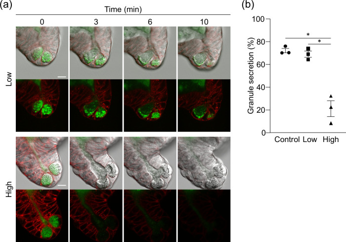
Fig 4. Inhibition of Paneth cell granule secretion by light illumination.
(a) The representative fluorescent and DIC images of Paneth cells acquired by low or high-dose light illumination for 10 min after adding 10 μM CCh. Paneth cell granules and cell membrane were stained by Zinpyr-1 (green) and CellMask (red), respectively. Scale bars: 10 μm. (b) Percent granule secretion 10 min after adding CCh. The values were depicted as mean ± standard error of the mean for three independent experiments. *P < 0.05.

