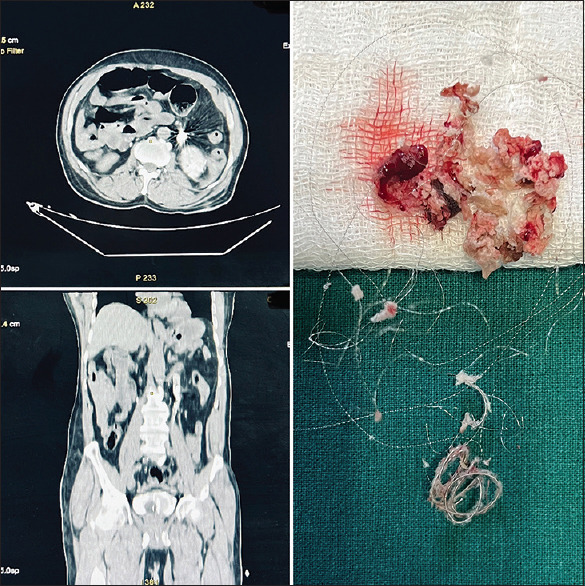
Figure 2.

(Left up) Computed tomography (CT) axial image showing hyperdense coil causing pelviureteric junction (PUJ) obstruction, (left down) CT Coronal section showing migrated coil at the PUJ. (right) Retrieved specimen of endovascular coil and glue
