FIGURE 5.

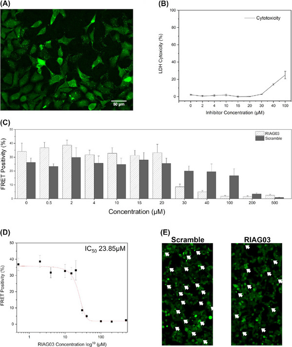
Lead peptide RI‐AG03 penetrates cells and reduces the seeding capacity of Tau seeds. (A) Fluorescence microscopy image depicting cellular uptake of 15 µM 5(6)‐carboxyfluorescein tagged RI‐AG03 [FAM‐RI‐AG03] by HEK‐293 cells incubated in DMEM/10% FBS at 37°C, with 5% CO2, after 24 h. Cells were seeded to a 96‐well plate at 20,000 cells/100 µL per well and supplemented with FAM‐RI‐AG03. Cells were visualized on a Nikon Eclipse Ti fluorescent microscope using the FITC filter. (B) LDH cytotoxicity assay using varying concentrations of RI‐AG03 co‐incubated with HEK‐293 cells. LDH Cytotoxicity (%) denotes lysed cells. Cells were seeded to a 96‐well plate at 10,000 cells/100 µL per well. Toxicity begins to increase at 40 µM. Experiments were conducted in triplicate. (C) FRET positivity indicative of Tau aggregation in a HEK‐293 biosensor cell line stably expressing Tau repeat domains with the P301S mutation fused to Cer/Clo. Cells were exposed to preformed Tau2N4R fibril seeds that were preincubated with either RI‐AG03 or Scramble peptide for 16 h before transduction. (D) Seeded aggregation is inhibited by RI‐AG03 at doses between 20 and 100 mM, with the IC50 being 23.85 mM. (E) Fewer green puncta indicative of Tau aggregates are seen in representative fluorescence microscopy images of the biosensor cells after treatment with preformed fibrils incubated with 30 µM RI‐AG03 compared with those incubated with Scramble peptide.
