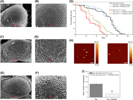
FIGURE 6.

Lead peptide RI‐AG03 reduces tau aggregation and suppresses phenotypes in a model of tauopathy. (A–F) SEM images of the eyes of drosophila melanogaster at 100 (A, C, E) and 50 (B, D, F) microns from left to right; N = 6 (A, B) healthy GMR‐GAL4 (C, D) GMR/hTau2N4R flies without treatment (E, F) GMR‐hTau2N4R flies treated with RI‐AG03 20 µM. (G) Survival curves of control (Elav/Orer) and Tau overexpressing flies (Elav/hTau2N4R) treated with low (0.08 µM) and high (0.8 µM) doses of the RI‐AG03, respectively, and no treatment. Log‐Rank test n = 100 per genotype/treatment group, p = 0.0007 and 0.0004, respectively. (H and I) Visualization (H) and quantitation (I) of reduction of Tau aggregates after 0.08 µM inhibitor treatment using atomic force microscopy imaging on an insoluble Tau prep obtained from 6‐week transgenic flies expressing hTau2N4R pan‐neurally. Without RIA03 treatment there is evidence of fibrils (arrow) and oligomers (arrowhead). After treatment, no fibrils are seen, only large spherical structures. n = 3, Unpaired t‐test p = 0.0045.
