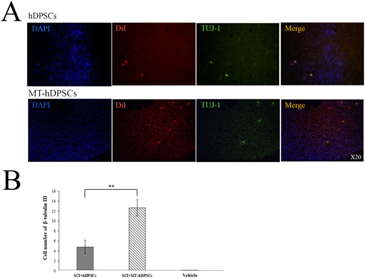
Fig. 6.
Differentiation of hDPSCs and MT-hDPSCs into neurons assessed by β-Tubulin III (TUJ-1) staining. (A) At the site of injury, the nuclei of hDPSCs and MT-hDPSCs were labeled with DAPI, the cells themselves were labeled with DiI, and their neuron differentiation was labeled with β-tubulin III. (B) A comparison of the number of β-tubulin III-positive cells among the Vehicle (SCI + DMEM), SCI + hDPSCs, and SCI + MT-hDPSCs groups demonstrated that MT-hDPSCs differentiated into neurons significantly more than hDPSCs at the SCI site (**p < 0.01). hDPSCs human dental pulp stem cells, MT melatonin, SCI spinal cord injury; magnification: ×20.

