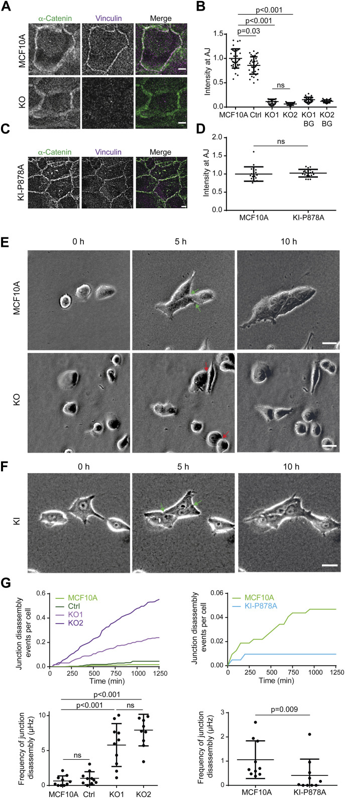
Figure 5. Vinculin stabilizes cell–cell junctions.
(A, B, C, D) Presence of vinculin at cell–cell junctions in KO and KI-P878A cells. (A, B, C, D) Staining of vinculin and α-catenin (A, C) and quantification of intensity at cell–cell junctions (B, D). Max z-projection from confocal microscopy. Scale bar: 5 μm. (B, D) Mean ± SD, n = 37 for (B) and n = 20 for (D), t test. N = 3 with similar results; pooled measurements from three independent repeats are plotted. (E, F) Time-lapse imaging of KO (E) and KI-P878A (F) cells in 3D collagen gels by phase contrast. Green arrows point at cell–cell junctions that were present at 5 h and that did not disassemble at 10 h, and red arrows point at cell–cell junctions that were present at 5 h and that were disassembled at 10 h. (A, B) Scale bars: 50 μm in (A); 25 μm in (B). (G) Quantification of cell–cell junction disassembly events per cell and frequency of cell–cell junction disassembly. Mean ± SD, n = 10, t test. N = 3 with similar results; pooled measurements from three independent repeats are plotted. Ctrl refers to the MCF10A cell line that has been genome-edited using a Ctrl gRNA.

