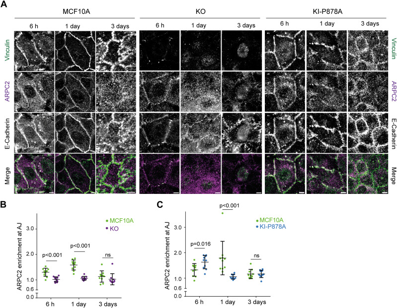
Figure 7. Vinculin retains Arp2/3 at cell–cell junctions.
(A) Staining of vinculin, ARPC2, and E-cadherin 6 h, 1 d, or 3 d after plating. Scale bar: 5 μm. Max z-projection from confocal microscopy. (B, C) Quantification of ARPC2 enrichment at adherens junctions in KO (B) and KI-P878A (C) cells. Mean ± SD, n = 10, t test. N = 3; 1 representative experiment shown.

