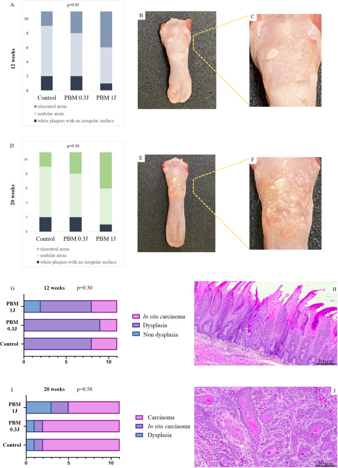
Fig. 2.
Clinical and histopathological features of our sample. (A), distribution of the cases according to clinical presentation in the 12 weeks; (B and C), white plaques with an irregular surface of one case at the end of 12 weeks; (D), distribution of the cases according to clinical presentation in the 20 weeks; (E) and (F), nodular areas with erythematous surface of one case at the end of 20 weeks; (G), distribution of the cases according to histopathological diagnosis in the 12 weeks; (H), epithelial dysplasia; (I), distribution of the cases according to histopathological diagnosis in the 20 weeks; (J), squamous cell carcinoma.

