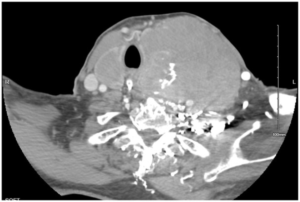
Image 1.

CT of the neck soft tissue with contrast: a large solid-appearing left neck mass with internal calcifications that appears similar in size up to 9.0 × 9.2 × 11 cm in size; this lesion appears to be centered within the left thyroid gland and extends into the superior mediastinum. There is a cystic nodule seen within the right thyroid lobe measuring 3.3 × 2.5 × 4.5 cm.
