Figure 2.

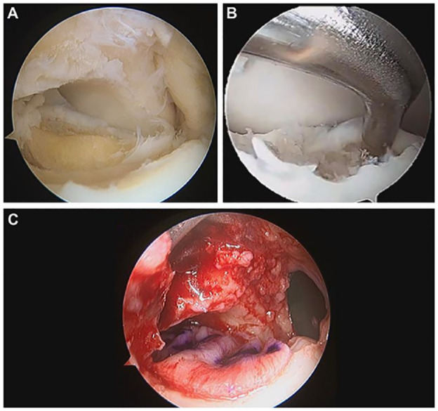
(A) Demonstrates an OLT after a curettage of the damaged cartilage. (B) Displays the intraoperative measurement of the lesion with an arthroscopic probe. (C) Shows the membrane placement over the OLT.

(A) Demonstrates an OLT after a curettage of the damaged cartilage. (B) Displays the intraoperative measurement of the lesion with an arthroscopic probe. (C) Shows the membrane placement over the OLT.