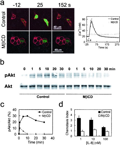
FIG. 5.
MβCD treatment impairs IL-8-triggered Ca2+ response and Akt phosphorylation and chemotaxis in response to IL-8 gradient. (a) IL-8 failed to induce CXCR1-CFP-mediated Ca2+ response in the MβCD-treated cells. CX1-HEK cells labeled with Fluo-4 (green) were stimulated with 50 nM of IL-8. Non-MβCD-treated cells (control) and MβCD-treated cells are shown. Upon IL-8 stimulation, changes in the intracellular Ca2+ concentration are shown in the graph on the right. (b) IL-8 stimulation induced CXCR1-CFP-mediated Akt phosphorylation in control cells but not in MβCD-treated cells. CX1-HEK cells were stimulated with IL-8 and subjected to Western blotting with antibodies for Akt and the phosphorylated form of Akt (pAkt). (c) Quantification of IL-8-induced Akt phosphorylation is shown. (d) Effect of MβCD on chemotaxis. CX1-HEK cell migration across 10-mm-pore-size filter membranes was measured in response to gradient of IL-8 of three different concentrations. In the absence of IL-8, the chemotaxis index of CX1-HEK cells is 1.

